 The Real Meal Revolution we run at Taylormade Performance & Rehabilitation is designed to help you lose weight, but the benefits pile up far beyond just looking a little better.Eating real food and ‘Feeling Great’ is a result of numerous ‘side-effects’ or ‘side benefits’ as Georgia Ede MD once described in a podcast with Ivor Cummins, when you use ‘real food’ to improve mental health rather than medications and their harmful ‘side effects’. Here is the 20min discussion for a further understanding of what she described. https://podcasts.apple.com/gb/podcast/the-fat-emperor-podcast/id1453181214?i=1000464487091
So the Real Meal Revolution program has shown benefits with its clients such as improved sleep, lowered blood pressure, remission of diabetes, clearer skin, reduced bloating, improved metabolism, boundless energy and improved blood readings. Boundless energy is something we all crave whether that is to be more productive at work, enjoying and being spontaneous with your partner or being able to keep up with your children. Health is essentially your most important goal because you want to live life fully, optimally and be the best you can be! So how can you test to see where you are right now? Perhaps you may know that something is just not right.‘ I don’t have the energy, my sleep is disrupted, my weight is going in the wrong direction, I am hungry all the time.’ When you’re gaining weight it is an external manifestation that something in the body is not right internally. So where do you start you may ask? Well it is fairly simple you need to take some measures some externally and some internally i.e a blood tests. There is a saying “if you don’t look, it don’t get fixed”. Or “you can’t change something until you know what you have been doing is wrong.” So what if you had some feedback beyond just a feeling something is not quite right? What do I look for? Markers of metabolic syndrome-is a clustering of at least three of the five following medical conditions, central obesity (abdominal fat-waist circumference measurement), high blood pressure, high blood sugar, high serum triglycerides, and low serum high-density lipoprotein (HDL). However the very best measure would be a post prandial assessment of insulin and glucose by an oral glucose tolerance test which will provide you with the best information from the root cause of the above symptoms -Hyperinsulinemia and insulin resistance. What are the guideline measures to know what is good? Good Markers are Waist circumference below 94cm M, below 84 FWaist circumference to height ratio below 0.5 [waist measurement (cm) at the belly button divided by height (cm)], Blood pressure below 125/75mmHg, Blood fasting glucose concentration below 5.0mmol/L, Blood Triglyceride concentration below 0.75mmol/L, Blood HDL cholesterol concentration above 2.0mmol/L. Disclaimer-These measures are from the Real Meal Revolution international edition. Please note that these measures should not be considered medical advice and exists for the purpose of sharing information only. These markers are associated with cardiovascular disease, type 2 diabetes, increased cancer rates, stroke, and links to Alzheimer’s which is being termed now as ‘type 3 diabetes’ as it follows the similar pathology as type 2 diabetes but is affecting the brain rather than the cardiovascular system. Now the increase in all these modern diseases may cause you wonder why. A pro-inflammatory diet has been very much the researched culprit (high in sugar, refined grains, trans fats, omega 6, food additives and sweeteners) essentially processed foods. So what to do? Once you have your baseline test results you can go to work on reversing your condition and improving the markers discussed above. First eat real foods get away from the pro-inflammatory diet and make your way towards being the best you can be or for a start and work everyday at being better than you were yesterday! If you would like to learn more and join the real meal revolution programme we run at Taylormade Performance & Rehabilitation please follow this link https://www.taylormaderehab.com/wellness-questionnaire.html fill in the survey and lets get you to live life to the full! About the Real Meal Revolution: Born out of South Africa and the Banting diet the Real Meal Revolution brought together Professor Tim Noakes ‘A1 rated scientist’ (A1 rating to a researcher who is “recognised by all reviewers as a leading scholar in his/her field internationally for the high quality and wide impact (i.e. beyond a narrow field of specialisation) of his/her recent outputs”) bestselling nutritionist Sally Ann Creed, and Top Ten rated restaurant chef Jonno Proudfoot is now changing lives all over the world with diabetes remission and other blood markers we may not even know are an issue. Eat Better South Africa Facebook page and http://www.thenoakesfoundation.org will provide you with further information for your interest. here to edit.
0 Comments
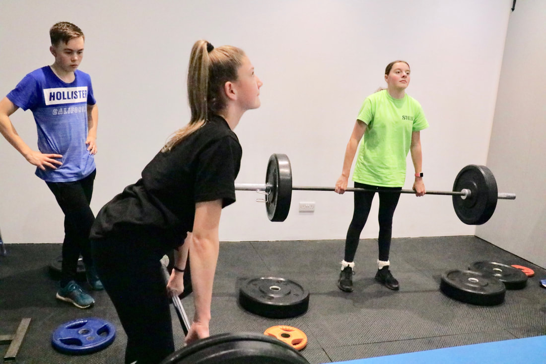 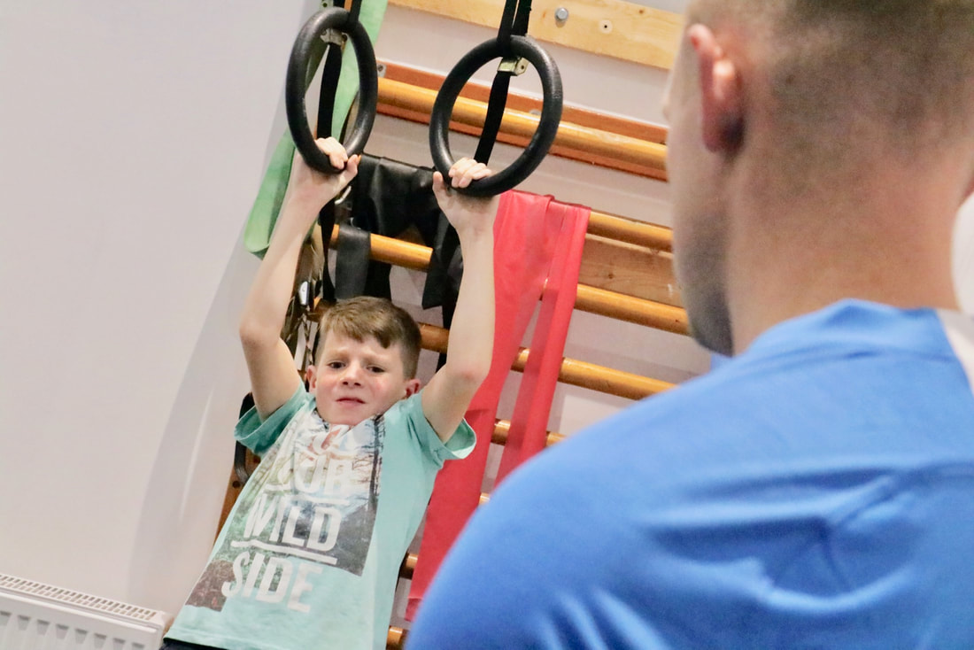 As parents and caregivers, we all want the best for our children's health and well-being. While physical activity is often encouraged for its numerous benefits, strength training sometimes takes a backseat when it comes to children and adolescents. However, recent research suggests that incorporating age-appropriate strength training into their routine can have significant advantages. Let's delve into the science-backed benefits of strength training for our young ones.
1. Enhances Muscular Strength and Bone Health: Contrary to common misconceptions, strength training does not stunt growth in children and adolescents. In fact, it can help build stronger muscles and improve bone density. The American Academy of Pediatrics (AAP) recommends that children as young as 7 or 8 years old can safely begin strength training with proper supervision and appropriate exercises. Regular strength training can lead to increased muscle strength, which can support better athletic performance and reduce the risk of injuries. 2. Promotes Healthy Weight Management: Childhood obesity is a growing concern worldwide. Strength training, when combined with cardiovascular exercise and a balanced diet, can support healthy weight management in children and adolescents. A study published in the Journal of Pediatric Obesity found that strength training exercises, alongside aerobic activities, significantly reduced body fat percentage and improved body composition in overweight and obese children. 3. Boosts Cognitive Function and Academic Performance: Physical activity has long been linked to improved cognitive function, and strength training is no exception. Research published in the Journal of Sports Sciences suggests that regular strength training can positively impact executive functions, such as attention, memory, and problem-solving abilities. These cognitive benefits can extend to the classroom, leading to improved academic performance. 4. Enhances Mental Well-being and Self-esteem: Strength training has been found to have a positive impact on mental health and self-esteem in children and adolescents. Engaging in strength training can boost confidence as children witness their own physical progress and achievements. Additionally, exercise releases endorphins, which are known to elevate mood and reduce symptoms of anxiety and depression. 5. Fosters Healthy Habits and Long-term Fitness: Encouraging children to engage in strength training at an early age sets the foundation for a lifetime of healthy habits. By introducing strength training as part of their regular routine, children are more likely to continue exercising into adulthood, reducing the risk of sedentary lifestyles and associated health issues. Conclusion: The benefits of strength training for children and adolescents are supported by scientific literature. Incorporating age-appropriate strength training exercises into their routine can enhance muscular strength, promote healthy weight management, boost cognitive function, improve mental well-being, and foster long-term fitness habits. However, it is crucial to ensure proper supervision, appropriate exercises, and gradual progression to minimize the risk of injury. By empowering our young ones with the knowledge and tools for strength training, we are nurturing their physical and mental well-being for years to come. Certainly! Here are some references to scientific studies on the benefits of strength training for children and adolescents: 1. Faigenbaum, A. D., & Myer, G. D. (2010). Resistance training among young athletes: safety, efficacy and injury prevention effects. British Journal of Sports Medicine, 44(1), 56-63. 2. Faigenbaum, A. D., & Myer, G. D. (2012). Pediatric resistance training: Benefits, concerns, and program design considerations. Current Sports Medicine Reports, 11(3), 161-168. 3. Behm, D. G., Faigenbaum, A. D., Falk, B., & Klentrou, P. (2008). Canadian Society for Exercise Physiology position paper: resistance training in children and adolescents. Applied Physiology, Nutrition, and Metabolism, 33(3), 547-561. 4. Lloyd, R. S., Faigenbaum, A. D., Stone, M. H., Oliver, J. L., Jeffreys, I., Moody, J. A., ... & Myer, G. D. (2014). Position statement on youth resistance training: the 2014 International Consensus. British Journal of Sports Medicine, 48(7), 498-505. 5. Moran, J., Sandercock, G., Ramírez-Campillo, R., & Clark, C. C. (2017). Effects of resistance training on pediatric obesity outcomes: a systematic review and meta-analysis. Sports Medicine, 47(6), 1261-1277. 6. Myer, G. D., Faigenbaum, A. D., Edwards, N. M., Clark, J. F., Best, T. M., & Sallis, R. E. (2015). Sixty minutes of what? A developing brain perspective for activating children with an integrative exercise approach. British Journal of Sports Medicine, 49(23), 1510-1516.  Hey there, fellow athletes! Have you ever wondered how some athletes seem to have that extra edge when it comes to performance? Well, one crucial factor that plays a significant role is metabolic health. In this blog post, we'll explore why getting a metabolic health check is so important for optimizing your performance and taking your athletic abilities to the next level!
Understanding Metabolic Health: Metabolic health refers to how efficiently your body converts food into energy and utilizes it during physical activities. It's like having a well-oiled machine that powers your muscles, fuels your workouts, and helps you excel in your chosen sport. By keeping your metabolic health in check, you can unlock your full potential as an athlete. The Importance for Athletes: 1. Tailoring Training and Nutrition: A metabolic health check provides valuable insights into how your body processes energy. It can reveal information about your calorie-burning rate, oxygen delivery to muscles, and overall energy utilisation. Armed with this knowledge, you can work closely with your coaches, trainers, and nutritionists to customise your training and dietary plans. This ensures that you're fueling your body optimally, making the most of your efforts, and achieving peak performance. 2. Identifying Performance Barriers: Ever experienced fatigue or hitting a plateau despite putting in the effort? Sometimes, underlying metabolic issues can hinder your progress. By undergoing a metabolic health check, you can identify any potential barriers to optimal performance. This might include issues such as insulin resistance, inadequate nutrient absorption, or inefficient energy utilisation. Armed with this knowledge, you can take proactive steps to address these challenges and elevate your performance. 3. Enhancing Recovery and Injury Prevention: As athletes, we push our bodies to the limits, which requires proper recovery and injury prevention. A metabolic health check can provide insights into how effectively your body recovers and replenishes energy stores. By understanding your body's unique recovery needs, you can design a personalised recovery plan that includes rest, nutrition, hydration, and targeted therapies. This helps you bounce back faster, reduce the risk of injuries, and maintain consistent performance over time. 4. Optimising Long-Term Health: While performance is crucial, long-term health should always be a priority. Metabolic health checks allow you to assess important markers like insulin, blood sugar levels, cholesterol, and other metabolic indicators. By monitoring these factors, you can proactively address any imbalances or early signs of metabolic disorders. This not only supports your athletic performance but also promotes overall well-being and longevity. Conclusion: Dear athletes, investing in your metabolic health is a game-changer. By getting regular metabolic health checks, you gain valuable insights into your body's energy utilisation, which allows you to fine-tune your training, optimise recovery, and prevent potential barriers to performance. Remember, your body is your most valuable asset on your athletic journey. Prioritising metabolic health ensures that you're equipped to unleash your full potential, both on and off the field. Keep striving, keep excelling, and keep pushing the boundaries of your athletic prowess! 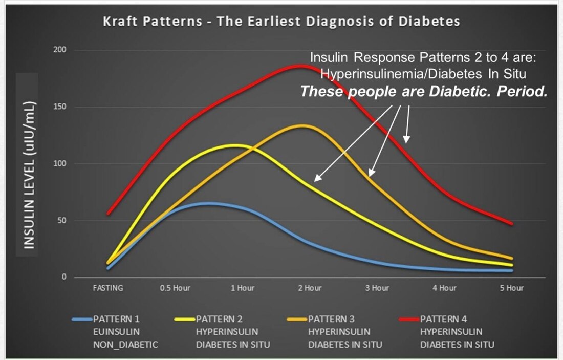 Did you know that there's a special test called the Kraft Test that helps you check if your body is using insulin properly? Insulin is a hormone that helps control the sugar in our blood. If our bodies don't use insulin well, it can cause problems like high blood sugar. But don't worry, there's a way to improve our health by eating real foods! What is the Kraft Test? The Kraft Test is a special test that to check if our bodies are using insulin properly. It's like a check-up for our insulin! You can do this test by checking your blood sugar levels before and after we drink 75g of glucose. Insulin is also measured to see how much insulin is needed to control the blood sugar levels. It helps you find out if you have something called insulin resistance, which means our bodies have trouble using insulin. Why is the Kraft Test important? The Kraft Test is important because it helps you find out if you are at risk for getting sick. If we have insulin resistance, we might be more likely to have problems like diabetes. Diabetes is a condition where our blood sugar levels are too high. By doing the Kraft Test, you can catch these problems early and help you stay healthy. Eating Healthy and Therapeutic Carbohydrate Restriction Now, let's talk about how we can improve our health by eating healthy foods! One way to do this is through therapeutic carbohydrate restriction. Carbohydrates are a type of food that gives us energy, like bread, rice, and pasta but these can increase our blood sugar just same as other carbohydrates, like sugary snacks and drinks. That's where therapeutic carbohydrate restriction comes in! It means eating fewer carbohydrates and prioritising protein and fats at your meals. 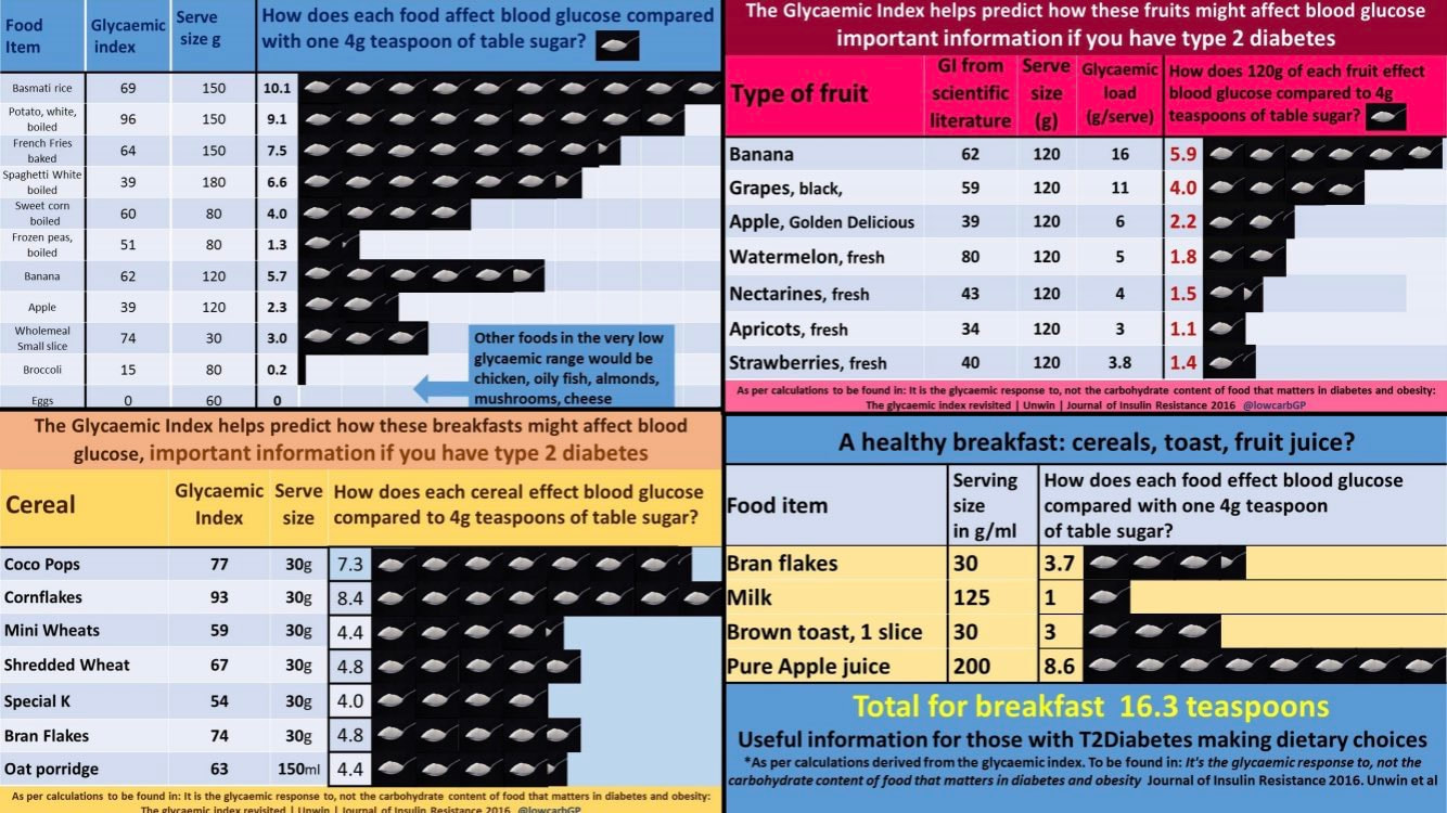 Tips for Eating Healthy
Here are some tips for eating healthy and improving our health through therapeutic carbohydrate restriction: 1. Prioritise protein and don't fear healthy fats 2. Eat fruits and vegetables: choose Fruits and vegetables that are packed with vitamins and minerals but also low in sugar, so they won't make our blood sugar levels go up too much. 3. Drink water: Water is the best thing to drink to stay hydrated. Sugary drinks like soda and juice can have a lot of sugar, which can be bad for our blood sugar levels. You can find mouthwatering recipes here a at https://www.dietdoctor.com/low-carb/recipes In conclusion, the Kraft Test is an important test that helps you check if your body is using insulin properly. By eating healthy foods and following therapeutic carbohydrate restriction, we can improve our health and lower our risk of getting sick. Remember, it's always a good idea to talk to a doctor or a registered dietitian for personalized advice on how to eat healthy and take care of our bodies. 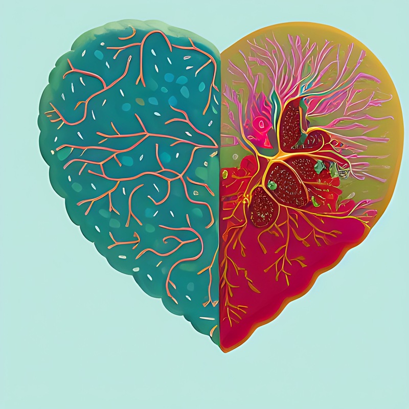 In today's fast-paced world, chronic diseases have become increasingly prevalent. Conditions such as Alzheimer's, high blood pressure, osteoarthritis, insulin resistance, type 2 diabetes, chronic stress, and lack of sleep are all interconnected and influenced by various lifestyle factors. We explore here the impact of lifestyle choices, specifically focusing on processed carbohydrates, on the development and management of these chronic diseases.
1. Alzheimer's: Alzheimer's disease is a neurodegenerative disorder characterized by cognitive decline and memory loss. While the exact cause of Alzheimer's is not yet fully understood, research suggests that lifestyle factors play a significant role. Chronic consumption of processed carbohydrates, such as sugary snacks and refined grains, has been linked to increased inflammation and oxidative stress in the brain, which may contribute to the development of Alzheimer's disease. 2. High Blood Pressure: High blood pressure, or hypertension, is a common chronic condition that affects millions of people worldwide. Unhealthy dietary choices, including excessive consumption of processed carbohydrates, have been associated with elevated blood pressure levels. Processed carbohydrates often lack essential nutrients and fiber, leading to weight gain and insulin resistance, both of which contribute to increased blood pressure. 3. Osteoarthritis: Osteoarthritis is a degenerative joint disease characterized by the breakdown of cartilage. While age and genetics are significant risk factors, lifestyle choices also play a crucial role in its development. Processed carbohydrates, particularly those high in added sugars, lead to chronic inflammation and weight gain, which can exacerbate osteoarthritis symptoms and accelerate joint damage. 4. Insulin Resistance and Type 2 Diabetes: Insulin resistance is a condition in which cells become less responsive to insulin, leading to elevated blood sugar levels. The consumption of processed carbohydrates, especially those with a high glycemic index, can contribute to the development of insulin resistance and type 2 diabetes. These types of carbohydrates cause rapid spikes in blood sugar levels, straining the body's insulin production and leading to long-term metabolic disturbances. 5. Chronic Stress: Chronic stress has detrimental effects on overall health and has been linked to various chronic diseases. Stress triggers the release of stress hormones, such as cortisol, which can disrupt the body's metabolic balance. Stress-induced cravings for processed carbohydrates are common, as they provide temporary comfort. However, the excessive consumption of these carbohydrates further exacerbates the negative impact of stress on health and contributes to the development of chronic diseases. 6. Lack of Sleep: Sleep is essential for overall health and plays a vital role in maintaining proper bodily functions. Lack of quality sleep has been associated with an increased risk of chronic diseases, including those mentioned above. Poor sleep patterns can disrupt hormonal regulation, leading to imbalances that contribute to inflammation, insulin resistance, and other metabolic dysfunctions. Additionally, sleep deprivation often leads to increased cravings for processed carbohydrates, further exacerbating the negative effects on health. The prevalence of chronic diseases, such as Alzheimer's, high blood pressure, osteoarthritis, insulin resistance, type 2 diabetes, chronic stress, and lack of sleep, can be influenced by various lifestyle factors. Among these factors, the consumption of processed carbohydrates plays a significant role. By making conscious choices to reduce processed carbohydrate intake and adopting a balanced, nutrient-rich diet, individuals can mitigate the risk of chronic diseases and improve overall health and well-being. Additionally, incorporating stress management techniques and prioritizing quality sleep are vital steps towards maintaining a healthy lifestyle and preventing the onset of these chronic conditions.  In recent years, therapeutic carbohydrate restriction has gained significant attention as a dietary approach to promote and maintain overall health. This practice involves limiting the intake of carbohydrates, particularly refined sugars and processed grains. By adopting carbohydrate restriction, individuals can enhance their body's ability to regulate blood sugar levels, manage weight, and potentially improve various aspects of their health. This essay explores the therapeutic benefits of carbohydrate restriction and how it can help our bodies stay healthy.
1. Blood Sugar Regulation: Carbohydrate restriction plays a crucial role in regulating blood sugar levels. When carbohydrates are consumed, they are broken down into glucose, which stimulates the release of insulin. By reducing carbohydrate intake, individuals can prevent spikes in blood sugar levels, ultimately improving insulin sensitivity. This can be particularly beneficial for individuals with insulin resistance, metabolic syndrome, or type 2 diabetes. 2. Weight Management: Carbohydrate restriction has shown promise in promoting weight loss and maintaining a healthy weight. When we consume excessive carbohydrates, our bodies tend to store the excess glucose as fat. By limiting carbohydrate intake, especially refined sugars and processed grains, individuals can tap into their fat stores for energy, leading to weight loss. Furthermore, a low-carbohydrate diet often helps reduce appetite and control cravings, making it easier to adhere to a calorie-restricted diet. 3. Metabolic Health: Carbohydrate restriction has been found to improve various markers of metabolic health. Studies have demonstrated that this dietary approach can lead to a reduction in triglycerides, an increase in HDL (good) cholesterol levels, and a decrease in blood pressure. These improvements in metabolic parameters contribute to a reduced risk of cardiovascular diseases and other metabolic disorders. 4. Inflammation and Chronic Diseases: Chronic inflammation is linked to the development of various diseases, including heart disease, obesity, and type 2 diabetes. Carbohydrate restriction has been shown to reduce inflammation markers in the body. By minimizing the consumption of refined sugars and processed grains, individuals can mitigate the inflammatory response, potentially lowering their risk of chronic diseases. 5. Energy and Mental Clarity: Contrary to popular belief, our bodies can function efficiently without relying heavily on carbohydrates for energy. When carbohydrates are restricted, the body adapts to using fats as its primary fuel source, a state known as ketosis. Many individuals report increased energy levels and improved mental clarity when in ketosis. This may be particularly beneficial for those with neurological conditions such as epilepsy or neurodegenerative diseases. Therapeutic carbohydrate restriction offers numerous benefits for maintaining and promoting overall health. By regulating blood sugar levels, aiding weight management, improving metabolic health, reducing inflammation, and providing sustainable energy, this dietary approach has the potential to positively impact our well-being. However, it is essential to seek guidance from healthcare professionals, nutritionists or LCHF coach to ensure that a carbohydrate-restricted diet is followed correctly and meets individual nutritional needs. With proper implementation, therapeutic carbohydrate restriction can serve as a valuable tool in our pursuit of a healthy lifestyle. 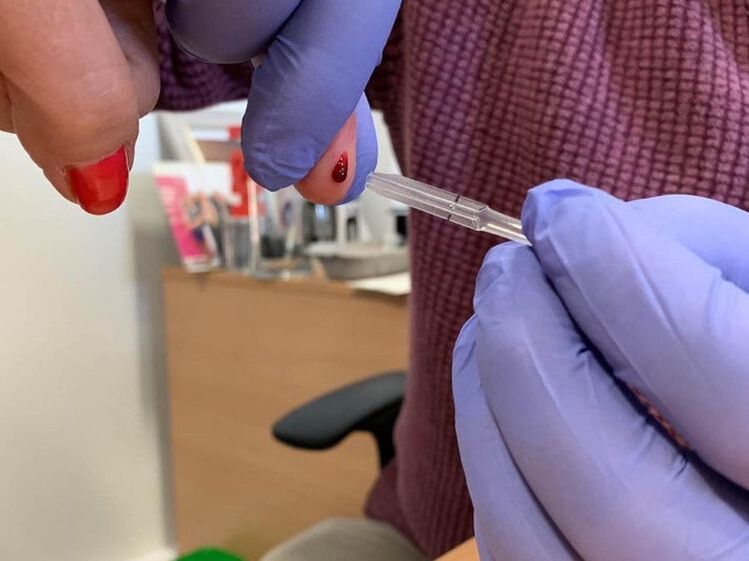  When it comes to staying healthy, something called metabolic health is super important. We also need to know about insulin testing. Let's learn why they matter so much!
1. Metabolic Health: Metabolic health means our body is working well and balanced. It involves things like blood pressure, cholesterol levels, blood sugar, and weight. When our body is metabolically healthy, we feel good, and it helps to prevent sickness. 2. The Role of Insulin: Insulin is a special hormone our body makes. It helps to control the sugar in our blood. It tells our cells to take in sugar and use it for energy. If our body has problems with insulin, we might get sick and have trouble managing our blood sugar. 3. Importance of Insulin Testing: a. Finding Problems Early: Insulin testing helps us find problems with insulin before they get really bad. This way, we can do things to stay healthy and prevent diseases like diabetes. b. Getting the Right Treatment: Insulin testing tells us how our body is using insulin. This helps us create a special plan just for us, like eating healthy foods and maybe bringing some more exercise into our lifestyle. c. Checking Progress: If we already have problems with insulin, testing helps us see if our treatment is working. We can make changes if needed to keep our blood sugar under control. d. Checking Overall Health: Insulin testing shows how well our body is handling sugar. It gives us a better idea of how healthy we are overall. Taking care of our metabolic health is super important for staying healthy and feeling good. Insulin testing helps us understand how well our body is using insulin and managing our blood sugar. It helps us find problems early, get the right treatment, and check on our progress. By paying attention to our metabolic health and getting insulin testing when needed, we can keep our bodies happy and healthy! 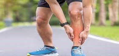 If you have spent any significant time with the running population, you will likely have heard of the term “Shin Splints”. This generic term frequently used to describe shin pain can have several underlying causes which all result in an annoying disruption of time spent running! This article will explore the main factors that can result in the over-arching label of Shin Splints and the different terminologies that may be discovered when investigating this condition. With some further understanding of the subtle difference in presentation and possible causes, appropriate early management of pain could be achieved. Medial Tibial Stress Syndrome 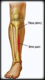 What is it? The medical world will more commonly describe exercise induced pain in the lower leg as ‘Medial Tibial Stress Syndrome’ (MTSS). This is thought to occur either when the muscles in the lower leg contract, causing a traction tension on the shin bone (Tibia) and tissue surrounding it (periosteum), or as a result of repetitive loading stress on the bone from impact activities. Its probably true that both schools of thought have relevance when considering the likely causes to this condition. What are the symptoms?
MTSS is classified as an overuse injury. It is commonly suffered among the running population due to the repeated impact stress to the lower limb experienced during this activity. Factors that can influence its prevalence are multiple and can be both intrinsic and extrinsic in nature. Intrinsic risks consider the biomechanics of the runner. Structural and postural inefficiencies such as low or high foot arches, the angle of the shin (tibial torsion or varum) or muscle imbalances can all have an impact on the stress on the muscle’s attachment to the bone. Extrinsically the risks are posed regarding sudden significant increases in running intensity (duration or frequency). Firm surfaces such as road running or hill running can cause additional stress. How to treat it
Stress fractures What is it? There are schools of thought that consider MTSS to be on a continuum, where the later stages present with a stress fracture to the Tibia. In essence a stress fracture is a repeated force creating stress on the bone at a rate greater than the bone can support. For a bone to adapt and become stronger it undergoes a degree of stress response. A healthy stress response causes microscopic degeneration of the bone which is sequentially reabsorbed creating a growth in the bone tissue. If this stress becomes more intense than the reabsorption rate, the microscopic degeneration increases and eventually a tiny crack in the bone can form. This is termed a stress fracture. If at this point the stress continues, the bone becomes at risk of a more significant fracture that is commonly recognised as a break. Tibial stress fractures are a common site. This bone is subjected to high impact weight bearing stress so is vulnerable when considering the activity of running. 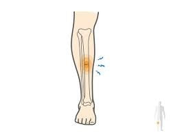 What are the symptoms?
How to treat it?
Anterior compartment Syndrome Chronic exertional compartment syndrome is something we have explored in a previous article www.taylormaderehab.com/blog/everything-you-need-to-know-about-calf-injuries-for-runners The lower leg is separated into 4 major compartments: the anterior, the superficial posterior, the deep posterior and the lateral compartment. The anterior compartment is in the front of the lower leg to the outside of the tibia. This syndrome can create symptoms that maybe mistaken for ‘shin splints’. 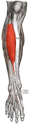 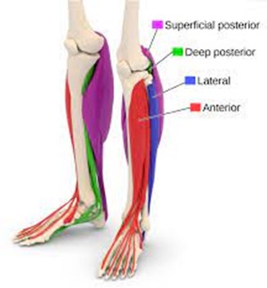 What are the symptoms?
A common site of pain that can take us off the road is in the lower leg known as the Calf. Perhaps the most obvious and familiar injury considered at the calf is a muscle strain. However, there can be some alternative culprits for this disturbance which are explored here.
Muscle Strain What is it ? Muscles are made up of many fibres, each fibre is programmed to shorten and lengthen (contract and relax). It is this contraction and relaxation mechanism that drives a joint to move. A strain of a muscle is when there is a tear in a portion of these fibres causing pain and a degree of functional deficit. What causes it? A muscle is most at risk of tearing when it is over stretched. This might be following a sudden movement like a sprint start, a jump or a slip. A muscle strain also has a greater risk of occurring if the muscle is fatigued or over-active. Symptoms The severity of a strain has three classifications: Grade I – is where a small portion of muscle fibres have been disrupted. This often presents as mildly painful. The muscle continues to function with no loss of strength. Grade II – Is where a moderate portion of the muscle fibres are torn. This presents as moderately painful. It can limit range, strength and function of the muscle. There may be evidence of swelling and bruising. Grade III – Is where all fibres are torn, resulting in a complete rupture. This is significantly painful and will result in a complete lack of function of that muscle. A pop or snapping sensation is often experienced. The muscle may look distorted with a large lump or depression where the muscle fibres have recoiled. This will be accompanied by significant bruising. How to treat it?
How to prevent it?
Chronic Compartment Syndrome What is it and what causes it? Major muscle groups are housed in a sheath of tissue known as ‘Fascia’ these create compartments and are found in the arms and legs. During activity, muscles increase in volume, ordinarily these compartments are large enough to accommodate this natural adaptation. However, occasionally the swelling creates an increase in pressure in the compartment causing discomfort. This is known as Chronic (exertional) compartment Syndrome. This condition can be experienced in the calf and can commonly affects both legs. Symptoms Symptoms are generally reported as a gradual onset of stiff, tight aching pain. This may include numbness, tingling, or change in sensation. Symptoms usually get worse as the activity continues and settle shortly after the activity has stopped. How to treat it?
How to prevent it?
Deep vein Thrombosis (DVT) What is it? A deep Vein Thrombosis is a blood clot in the deeper layers of veins. These will most often occur in the lower limb and pelvic area and are more prevalent in the calf. The danger of a DVT is that the clot which is initially lodged in the vein can break away and travel to the lungs risking the occurrence of a Pulmonary Embolism (PE). A PE is a serious condition as it can be potentially fatal. Symptoms A DVT can feel insignificant and can often be miss interpreted as a muscle strain or cramp. It often presents as a dull ache deep in the calf which gets worse with activity. There may be the experience of swelling, redness or discoloration to the affected limb. The area may feel warm to touch, tender or present with bruising. What causes it?
How to treat it?
How to prevent it?
Foot pain is common in runners for a number of reasons. The very nature of running makes it a high impact repetitive activity. The impact of that force is primarily experienced in the foot where mild imbalances will have a significant effect. The highly complex synergy between the muscles, ligaments and bones in the foot relies on these tissues working in harmony. Foot pain can be long standing and niggly and can often end up making it almost impossible to run. Identifying these injuries and their causes can be crucial to a pain free return to the open road! Here are some common foot injuries suffered by runners. Plantar Fasciitis 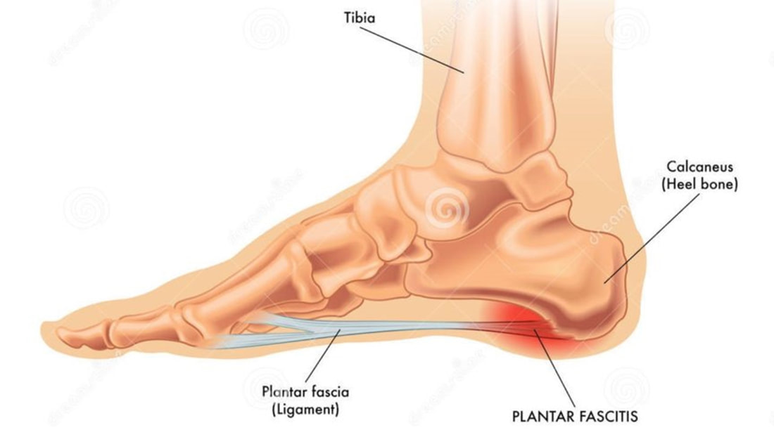 What is it and what causes it? The Plantar Fascia is a thick wide band of strong connective tissue. This ligament connects the heel bone (Calcaneus) to the base of the toes (Metatarsals). Its primary role is to support the arch of the foot helping to dissipate the load through the foot. Most commonly, Plantar Fasciitis is considered an overuse injury which occurs when a repetitive force is created that the plantar fascia can no longer support. Over time this can create a degeneration of the Plantar Fascia. Onset of pain may occur following an intense introduction to a new activity such as running or a sharp increase in training intensity. Predisposing factors to this injury might be excessive tightness in the calf and insufficiency in supportive footwear. A sharp pain is experienced in the sole of the foot close to the heel. The pain is usually at its worst following a prolonged period of rest, specifically first thing in the morning, which may ease slightly after a few steps. Symptoms may be aggravated once again following exercise or long periods of standing. How to treat it?
Stress Fracture 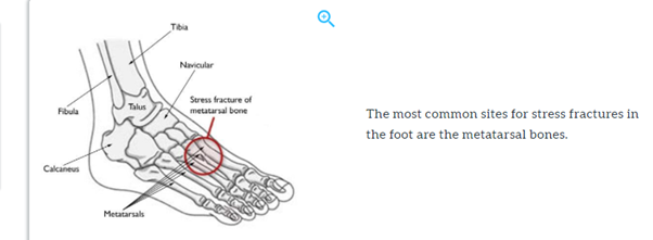 What is it and what causes it? A stress fracture is a small crack in the bone. It is so small it is often not even picked up on an x-ray. A stress fracture occurs along the continuum of a bones’ stress reaction. If caught early enough a stress reaction will present as a bone bruise, if symptoms are ignored or stress load is not adapted sufficiently then a stress fracture will develop. Pain on weightbearing is the most significant symptom. This is worse on impact and settles at rest. It is often a localised sharp pain pinpointed to the site of the fracture. Swelling on the top of the foot can be observed depending on the fracture’s location. The most common running related stress fractures in the foot are along the 2nd or 3rd Metatarsal. This is the site of greatest load as the foot moves through the push off phase of the running gait. Depending on foot mechanics, they can also be present in the Calcaneus, Talus or Navicular bone. Occurring as an overuse injury, a stress fracture will develop over a period of time. It is frequently reported to affect the lower limb in high impact repetitive activities such as running and jumping. A bone is at risk of a stress fracture when a sudden increase in training load and intensity is experienced. This may equate to increasing the number of running sessions in a week or running longer distances. Other factors causing a stress fracture may be a change in surface conditions from soft to hard, inappropriate, or unsupportive footwear or bone insufficiency. How to treat it?
How to prevent it?
Metatarsalgia and Morton’s Neuroma 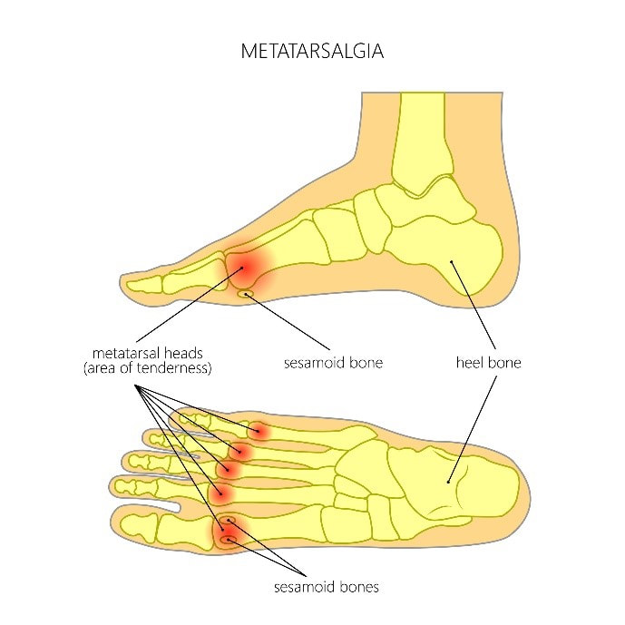 What is it and what causes it?
Metatarsalgia refers to inflammation to the heads of the Metatarsal bones (these bones sit in the forefoot between midfoot and toes). This occurs when there is a repetitive trauma to the sole of the foot such as the impact of running and jumping. Pain can develop suddenly or over a period of time and is experienced in the ball of the foot. This pain can be described as sharp, aching, or burning which often get worse with standing, walking or running. This condition can be similar to a Morton’s Neuroma where the tiny nerves between the toes are affected. Both can present with similar symptoms of pain and tingling or numbness. The most significant difference is in the site of pain. A Morton’s Neuroma usually affects the space between the 3rd and 4th toes where a thickening in tissues can be felt. This is often described as having a small pebble in the shoe. Other non-related running causal factors could be, excessive wearing of high heels, wearing of ill-fitting shoes, foot deformities such as bunions or hammer toe, obesity and diabetes. How to treat it?
If in doubt about any symptoms or pain does not settle, it can be most helpful to seek advice from a registered Health Care Practitioner to ensure that you have an accurate diagnosis and to ensure an effective treatment plan is prescribed.  A runner is as susceptible to ankle pain as any other lower limb injury. The repetitive demands of continual load acceptance during the running gait can create a high level of stress through this joint. The result ranges from an increasingly uncomfortable run to a complete inability to weight bear. Either scenario can be catastrophic to a carefully planned training programme or even the disruption to a regular health and wellbeing activity. The Ankle is made up of three bones, the Tibia and Fibular of the lower leg and the talus in the foot. These bones are held together by a series of ligaments most of which run along either side of the ankle joint. These ligaments offer stability to the ankle. There are also several tendons that cross the ankle attaching to muscles higher up the leg which create the movement of this joint. Each of these structures can become injured when running. The most common causes and types of injury are discussed in this article. Sprains What is it and what causes it? An ankle sprain is a generic term for an injured ligament in the ankle. However, there are numerous ligaments that can become injured in the ankle, all of which may be caused by a different movement. A sprain is caused when a joint is stretched passed its natural available range. Ligaments are bands of connective tissue creating an attachment between bones, forming a joint. When the joint is over-stretched a ligament becomes stressed to the point it can tear. The most common sprain is a lateral ankle sprain. It is often termed a ‘rolled ankle’ this affects the ligaments on the outside of the ankle. It is also possible to sprain the ligaments that support the inside (medial aspect) of the ankle too. Site of pain is usually a good indicator of which ligaments you have injured. The least common of ankle sprains is a high ankle sprain (Syndesmotic injury), this effects the connective tissue between the two lower leg bones (Tibia and Fibula) and commonly causes pain just above the front of the ankle joint as supposed to the outside of the joint as with a common lateral ankle sprain. Just like Muscle strains, ligament sprains can be classified in the three categories, grade I, II or III. 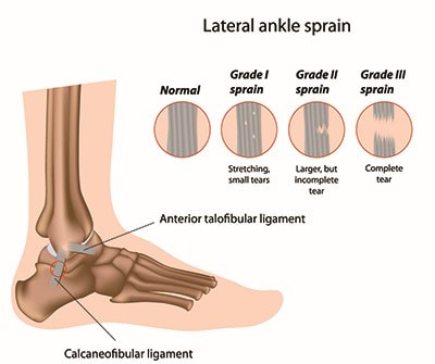 Runners may suffer an ankle sprain of any type by landing awkwardly into a stride, slipping off a curb, falling in a pothole, or tripping on uneven ground during a trail run. Symptoms
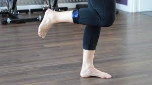 How to prevent it?
Stress fractures What is it and what causes it? Stress fractures are common in runners and have been widely discussed in the guide to running injury series. The repetitive high impact forces created during the running gait cycle are absorbed by many bones in the lower limb. Over time these repetitive forces result in microscopic trauma to the bones. Through inappropriate training programmes, this microscopic trauma does not have chance to remodel and can progress to create fine cracks (stress fractures). Most common stress fractures in the lower limb are found in the foot or shin. However, the Talus and Navicular bone are not immune to these stresses. They are located within the foot just below the ankle joint. Stress fractures will present with pain on weightbearing. This is worse on impact and settles at rest. It is often a localised sharp pain pinpointed to the site of the fracture. In the case of the talus and Navicular this may be in the front of the ankle or deep within the ankle joint. 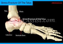 How to treat it?
How to prevent it?
Avulsion fractures What is it and what causes it? An avulsion fracture is when the forces stressed on a ligament or tendon are such that they create too much strain on the attachment causing a small piece of the bone to be pulled away with the ligament or tendon. The mechanism of these injuries is largely the same as a sprain and often presents in a similar way. Generally this is diagnosed by X-ray or CT scans. 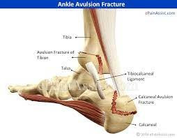 How is it treated?
Tendinopathy A tendinopathy is the degeneration of a tendon. Tendons attach a muscle to the bone. A tendinopathy occurs when a tendon is overused and subjected to adverse stresses. The subsequent degeneration is displayed as thickening, splitting or even tearing of the tendon. The repetitive nature of running and the high impact on the foot and ankle mean the numerous tendons around the ankle are vulnerable to tendinopathies if not managed sufficiently. Common sites affected are:
Symptoms associated with all these tendinopathies would be pain, swelling, weakness and even possible balance disturbances at the site of the respective tendon. As with all tendon injuries the pain tends to be prominent at the start of activities settling as the activity progresses. It may completely disappear at rest. Tendinopathies rarely get better without intervention and should not be ignored. 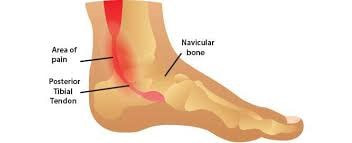 Tibialis posterior tendinopathy 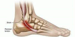 Peroneal tendinopathy 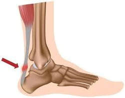 Achilles Tendinopathy How to treat it?
The hamstring is one of the most important muscle groups while running. It has a variety of roles throughout the gait cycle. These include forces to control the knee as it extends thorough the swing phase and creating the hip extension in the stance phase as the body passes over the planted foot. There is no wonder that this can be such a problematic region of the body when considering injuries that a runner might suffer. With such a complex role, pain in this muscle group can be caused by various injuries along the hamstring. This article will explore some of these most commonly suffered by runners. Hamstring Tear/Strain What is it and what causes it? Muscle strains are categorised into three grades. Generally, a hamstring strain will occur in the muscle belly (in the middle of the posterior thigh) or at either end of the muscle belly in the musculotendinous junction (as the muscle blends into the tendon). The most significant of strains (grade II or grade III) are more commonly experienced in sprinters due to the explosive forces. These types of strains can also be suffered because of sudden change of direction or acceleration and deceleration. Trail or fell runners who leap, jump or frequently run hills may be more susceptible to these types of strains than the steady state road pounding miles of a distance runner. The grade III rupture is a significant injury which will stop the runner in their tracks. It is often described as a pop or crack. The runner will feel like they have been shot in the back of the leg and is likely to leave them lying on the floor in severe pain. There will be immediate bruising and swelling as the blood rushes through the torn muscle. There will be a reluctance to put weight through this leg and the runner will be unlikely to walk. This would need immediate medical attention and, in most instances, result in surgical repair. A grade II strain will present similarly to a lesser extent. Although painful the runner would likely be able to walk, and subsequent bruising and swelling may be less severe or even non-existent. The more commonly experienced hamstring strains for steady state distance runners are the low grade I strains. The principle of injury is the same. The muscle is overloaded to the point it becomes under stress enough to tear. The over loaded muscle could have been caused by fatigue with increased intensity or lack of recovery in training. The tight shortened muscle in this state can create increased tension through the fibres as the hamstring goes through the gait cycle. These forces can expose the fibres to tear. A grade I strain will cause the runner a degree of pain but perhaps something that they can continue to run through. This pain usually stops after a run leading the runner to believe there is not an injury present. There is also unlikely to be any visible bruising or swelling present. 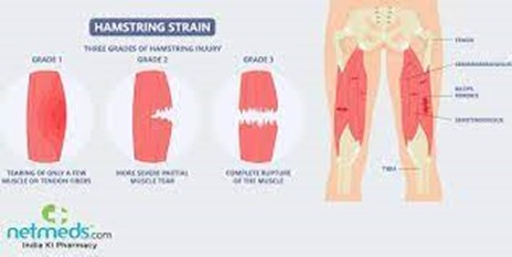 How to treat it?
How to prevent it?
Tendinopathy 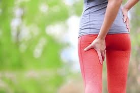 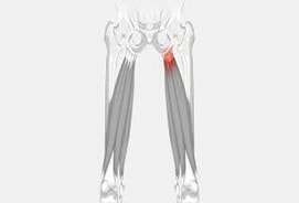 What is it and what causes it?
Proximal Hamstring tendinopathy is regarded as a commonly acquired injury in longer distance steady state runners. This is due to the gradual onset produced over time with a repetitive gait cycle. As previously discussed, the Hamstring muscle is under tension across two joints in the later stages of the swing phase (as the knee decelerates through extension and the hip drives into flexion). This position is thought to generate compression of the tendon at the top of the hamstring, causing irritation, inflammation, and pain. Aggravating factors may be a sharp increase in volume or intensity of running, an increase in stride length, or swift introduction to interval/fartlek training or hill running. Symptoms
Common knee injuries experienced by runners
Generally, people run to gain a positive reward, whether that is to improve wellbeing and achieve a physically active lifestyle or to achieve an ambition of running a Marathon or just simply because they enjoy the space and freedom. If this positive reward is met by frustrating challenges such as pain and injury it can soon become a negative experience. There are times when activities such as running can seem to bring about injury. There is usually an explanation for this and therefore a solution! Here are some common knee injuries suffered by runners. Patellofemoral pain syndrome (runner’s knee) What is it and what causes it? This is a common Injury suffered by Runners. It is often termed 'Runner's Knee'. In these circumstances, this type of Knee pain often comes on gradually over time. It can cause swelling in the Knee and generally presents as a dull ache in the Knee or behind the Knee cap. It is aggravated by activities such as walking, running and going up and down stairs. Common causes of this type of injury in runners are thought to be overuse and misalignment of the knee. There are many reasons for misalignment of the knee and it would be helpful to establish which of these is involved when addressing future prevention strategies. How to treat it?
Iliotibial band syndrome (ITBS) What is it and what causes it? Pain is experienced on the outside of the knee, there may be swelling present here. The pain is felt during running, usually coming on after a short while of activity and subsiding once activity has ceased. Often this is seen in longer distance runners and is exacerbated by running downhill and downstairs. ITBS is generally classed as an overuse injury where the tissue of the Iliotibial band (fascia running down the outside of the leg) is experiencing friction over the femoral condyle (bony surface) underneath. How to treat it?
Patella tendonitis What is it and what causes it? This is an inflammation of the tendon over the patella. The most commonly experienced site of pain is just below the patella as the tendon joins the Tibia. It is known as another overuse injury causes by repeated impact. In the early stages of this injury pain may only be felt at the start of a run and ease as the runner warms up. Perhaps becoming stiff once again following the end of the run. If left this may become more symptomatic throughout the whole of the run along with affecting daily living activities such as going up stairs or standing from a seated position. How to treat it?
Ligament injury What is it and what causes it? There are four main ligaments in the knee. Too much stress on these ligaments can cause them to overstretch and even tear. Ligament injuries are often resultant of a specific trauma such as a sudden twist, an awkward landing, an over straightening or a direct impact to the knee. If damaged the knee will often experience sudden sharp pain and swelling. It will likely be painful to weight bear and there could be a degree of instability. How to treat it?
It can be most helpful to seek advice from a registered Health Care Practitioner to ensure that you have an accurate diagnosis and to ensure an effective treatment plan is prescribed. Low back pain can not only affect our enjoyment of running but it also affects most of our daily living activities. This is not a pain that should be ignored, there are most likely elements of your running practice that can help address this niggling pain and the subsequent physical and mental energy drain suffered throughout the rest of your day. The repetitive high impact forces associated with running puts additional load through the spine. If the spine is not supported efficiently by the structures around it, pain, instability and muscle over activity can occur. Here are a few common conditions which can be experienced by runners. Sciatica What is it and what causes it? Sciatica is a widely used term to generically describe an irritation or inflammation to the Sciatic nerve. This is a large nerve that originates from the central spinal column in the lower lumber spine. It tracks through the buttock and down the back of the leg. The irritation to this nerve is caused by an impingement (compression) from numerous possible structures. The most common are at the intervertebral discs or the vertebral joints in the spine. Another culprit to display sciatica symptoms is the Piriformis (a muscle in the buttock). Sciatica is classically described as pain in the lower back radiating into the buttock and down the rear of the leg. The pain can be described as shooting or burning and can also present with numbness or pins and needles. Severe cases can result in loss of feeling, strength or function and change in sensation to the lower limb radiating as far as the foot. This article will look further into each of these causing factors to Sciatica. 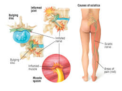 Intervertebral Disc herniation (bulge/prolapse) What is it and what causes it? One of the most common causes of Sciatica is a Disc herniation. Intervertebral discs are found between each vertebrae they act as a cushion throughout the spine offering support and protection. The disc are made up of a ‘jelly-like’ substance called the nucleus which is encased in a more firm elastic type substance known as the annular. A herniated disc is when some of the nucleus pushes out from the centre of the disc through a tear in the annular. This bulging nucleus can compress the nerve at its root causing the Sciatica symptoms. This type of injury is commonly the result of a degeneration of the disc. This aging disc becomes less flexible and prone to tearing. Modern lifestyle posture such as slouching or excessive loading of these joints can stress the integrity of the discs. The activity of running with its repetitive high impact loading in a pre-stressed spinal posture can increase the possibility of a disc herniation. Spinal stenosis or Spondylolisthesis What is it and what causes it? Both of these tend to cause lower back pain due to a degeneration in the spinal structures. A Stenosis is the narrowing of the spinal canal. The spinal canal is a space through the center of the vertebrae, which is where the central spinal cord travels. The narrowing can be the result of thickened degenerative ligaments or arthritic changes to the bone. This over laying of bone can narrow the spinal canal leading to compression of the spinal cord. This will present as pain in the back and often refers the sciatica symptoms described above. A Spondylolisthesis is an instability of the vertebrae, excessive movement can result in a slippage of one vertebrae onto another, this may be as a result of a fracture (more common in younger athletes) or from degenerative changes to the spine commonly seen in the over 50 population. The slippage of the vertebrae causes instability at this level of the spine and subsequent risk of compromising the space in the spinal canal. Symptoms would consist of pain in the lower back and into the buttocks, pain on walking or standing for long periods of time, pain on bending backwards. The impact of running particularly on firm surfaces is likely to aggravate symptoms of pain due to the repetitive high impact. How to treat these spinal conditions?
How to prevent them?
Piriformis Syndrome 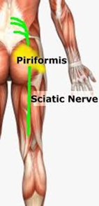 What is it and what causes it? Piriformis syndrome is a term used to identify the over activity of one of the deep buttock muscles (the Piriformis). This muscle is vital in the stability of the pelvis whilst running. When this muscle is overactive or fatigued it spasms causing pain. There can be instances of lower back pain associated with this condition. More commonly the pain radiates from the buttock on the affected side, tracking into the posterior thigh, creating the symptoms of sciatica described above. Depending on the individuals anatomy, occasionally the Sciatic nerve can become compressed by the Piriformis muscle. Most commonly when this muscle is overused, or training intensity is progressed too quickly. The Piriformis becomes tight and compresses the nerve within its muscle fibres. Pain can be experienced with prolonged sitting or standing from a seated position or while preforming a squat movement. This pain may ease with gentle movement. There may be tightness or restriction in the buttock on stretching it in a seated figure of four pose. There may also be pinpoint pain and tightness over the upper buttock to one side. How to treat it?
How to prevent it?
Sacroiliac Joint Dysfunction 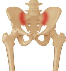 What is it and what causes it?
The sacroiliac joint is located at the joining of the pelvis to the sacrum (the base of the spine). Its main role is to accept the weight bearing load between the upper and lower body. This joint can become instable or restricted, both of which can result in pain in the lower back and buttock. If the neural structures are affected Sciatic like symptoms can be presented. A dull aching pain is often one sided and can be aggravated by weight bearing activities such as walking or running, sitting for a prolonged period, or standing from a seated position. Running can bring about an onset of pain due to the repetitive high impact loading particularly if this is on firm surfaces. The Sacroiliac joint can also become injured following a fall, over striding while running downhill or stumbling off a curb. How to treat it?
How to prevent it?
If in doubt about any symptoms or pain does not settle, it can be most helpful to seek advice from a registered Health Care Practitioner to ensure that you have an accurate diagnosis and an effective treatment plan is prescribed. A Kraft Test is a blood test used to identify a condition known as hyperinsulinemia. It is a picture of how your body responds to food, in particular glucose - the basic component of most of the carbohydrates we eat increasingly in our modern diets. When glucose enters the body it’s important that its concentration in our bloodstream is kept under control, so the body quickly responds to the glucose influx with a regulating hormone called *insulin*. Most of us associate insulin with a treatment for diabetes, and it is. But actually, everybody naturally produces insulin in the pancreas. Some of us need more insulin than others and the particular “pattern” of that response is very telling as to the status of your metabolic health. In fact, many medical professionals think it’s the most important blood test for metabolic health. The reason it is so important is because the test is an early-warning of chronic disease risk. A Kraft Test can reveal if the patient has a condition known as hyperinsulinemia, which is known to increase the risk of a host of conditions including diabetes but also hypertension, heart attack, stroke, neuropathy and even things like tinnitus, vertigo and hearing loss. In fact, Kraft took to calling hyperinsulinemia “occult diabetes” or “hidden diabetes”. In his original study*, Dr. Joseph Kraft, the doctor and pathologist who discovered these patterns, grouped the patterns he observed in patients into five broad groups. These patterns were observed after asking his patients to take a glucose drink while in a fasting state. This allowed him to isolate the behavior of insulin as it responds to glucose over the subsequent 3 hours and is almost exactly the same protocol you will follow to discover your Kraft Pattern. His discoveries were remarkable. He found that of patients who had normal blood glucose levels (about half the population in his study), and who therefore were thought to be in a “normal” or healthy metabolic state, 50% actually had hyperinsulinemia and a further 25% were borderline for the condition. Effectively these people had an early form of diabetes and didn’t even know it. They thought they were fine! The key insight a Kraft Test offers is in the pattern of insulin response to glucose consumption. But what do these patterns actually look like and what do they mean? Let’s take each one in turn. What follows are high-level descriptions and meaning of each Kraft Pattern as described by Crofts*. 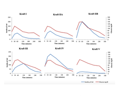 Pattern I
This is the pattern observed in normal metabolically healthy individuals. Note how the insulin responds to the consumption of glucose but does so in a moderate way and rapidly declines after it has done its job. Pattern IIA This pattern is observed in patients who have a “borderline” degree of insulin resistance. Here we see a higher fasting level of insulin (not a good sign) stronger initial insulin (post-glucose drink) concentration in the blood for the simple reason that more of it is needed to keep glucose under control. Pattern IIB This pattern is observed in patients who have what is called hyperinsulinemia - an early biomarker for diabetes risk. Here we once again see a higher fasting level of insulin. Importantly we also see an even stronger initial insulin (post- glucose drink) “spike” in the blood compared to those in Pattern IIA. Pattern III This pattern is also observed in patients who have hyperinsulinemia. Here again we see the characteristic higher fasting an post-glucose-drink insulin levels. But we also see a delayed “tail-off” in insulin as the hormone stays around longer in the blood. This is a more pronounced form of hyperinsulinemia and people with diabetes typically have this kind of pattern. Pattern IV A rarer pattern that some patients will present with. Here we see a very high fasting level of insulin, a non-delayed peak and then a long tail off. Again, this is likely to be a sign of diabetes. Pattern V Pattern V is actually a special case. Note how insulin stays very low throughout the test. This is because these individuals are likely Type 1 Diabetics. The opportunity to get this assessment is now and with new technology you can get a real time assessment at our clinic in Devon. *Detection of Diabetes Mellitus 'In Situ' (Occult Diabetes) - Joseph R. Kraft, M.D. Laboratory Medicine, Volume 6, Issue 2, 1 February 1975, Pages 10–22. Postprandial insulin assay as the earliest biomarker for diagnosing pre- diabetes, type 2 diabetes and increased cardiovascular risk - DiNicolantonio JJ, Bhutani J, OKeefe JH, et al. Open Heart 2017. Identifying hyperinsulinemia in the absence of impaired glucose tolerance: An examination of the Kraft database - Crofts C, Schofield G, Zinn C, Wheldon M, Kraft J. Diabetes Res Clin Pract. 2016 Aug. |
Taylormade
|

 RSS Feed

|
PERFORMANCE AND REHABILITATION CLINIC
Injury Management and Conditioning Specialist
Taylormade Rehab, 1 Belle Vue, Fenny Bridges, Honiton, Devon EX14 3BJ | 07800 824557 | 07855 386634
© 2019 Taylormade | Performance & Rehabilitation Clinic. Injury Management & Conditioning Specialists | Website design by brightblueC
© 2019 Taylormade | Performance & Rehabilitation Clinic. Injury Management & Conditioning Specialists | Website design by brightblueC